
Day 3 embryo cuture
From the moment of fertilization, the embryo’s first divisions have been the oocyte responsibility, as the DNA and proteins synthesis took place before fertilization. Therefore, day 3 of embryo culture represents the end of a phase in which the oocyte has been responsible for most of the embryonic development to give way to the entity of the embryo itself.
After day 3 of culture, gene expression will begin again, in this case, the embryonic genome.
The day 3 embryo evaluation allows us to establish an estimate of its potential to reach the blastocyst phase. To do this, we must visualize the embryos and evaluate the morphokinetic parameters. These parameters are the same as those evaluated on day 2 of embryo culture. Among them we can find:
- Number of cells and rate of division,
- Fragmentation degree,
- Number of cores,
- Changes in the zona pellucida and
- Cells shape and appearance (size, contour, cell cytoplasm, mottling, vacuoles, compaction).
Índice
Day 3 embryo rate de division
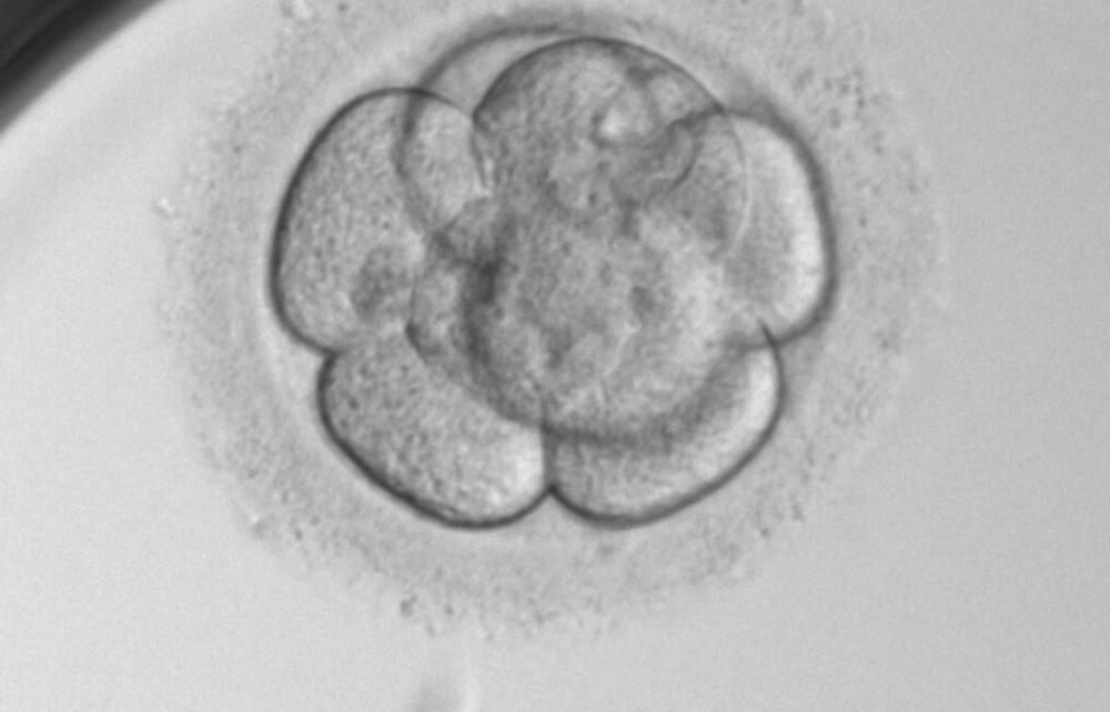
On day 3, the embryo division is expected to be between 6 and 8 cells, this will depend on the number of cells it had on day 2, which will provide us with information about the division rate. It’s possible to find embryos with faster development on day 3. There’s controversy in this regard about the potential of these embryos, although it has been proven that they reach the blastocyst phase in equal measure.

Cell stages of 6 to 8 cells

Cell stages of 9 and 10 cells
Embryo morphological characteristics on its third day of developement
The presence of cell fragments is common in human embryos and doesn’t always implies a low implantation rate, although it does have a negative effect on development. There’re 4 levels of fragmentation ranging from 1 to 4 depending on the embryo percentage they occupy:
- Grade 1: less than 10%
- Grade 2: 11-25%
- Grade 3: 26-35%
- Grade 4: over 35%

Fragmentation grade 1/Fragmentation grade 4
Other morphological parameters valued are shown in examples in the following images:

Cells asimetry /Multinucleation

Vacuoles / Zona pellucida, septate and oval
Day 3 embryo compaction degree
As already mentioned in the post, day 3 marks the end of one stage in the embryo development and the start of another, in fact, early compaction is a sign of embryo activation. A close intercellular contact is observed, even not being able to differentiate between one cell and another.
A: Pre-embryo without obvious signs of cell adhesion initiation.
B: Pre-embryo with signs of initiation of cell adhesion.
C: Compacted pre-embryo.
Pitting or mottled
Cytoplasmic mottling is a sign of cell cytoplasm activation, and just as compaction, it occurs during embryonic activation on day 3.

All these parameters are evaluated in the in vitro fertilization laboratory in order to establish which embryos are more likely to implant, establishing a graduation of A, B, C and D.
Dr Dori Rodríguez, biologist at Instituto Bernabeu.
IT MAY ALSO BE OF INTEREST TO YOU:
- Blastocyst embryo: What it is, advantages, types and classification according to its quality
- In Vitro Fertilization (IVF)
- Embryo transfer
- Criteria for embryo classification
- Embryonic arrest, why don’t all of my embryos develop equally?