What does embryo biopsy involve?
Preimplantation Genetic Diagnosis (PGD) is a tool designed to “get to know” the embryos genetically before they are transferred into the mother’s uterus. Thanks to this technique, we can study their chromosome count and find out if they are carriers of a hereditary disease. This information helps us to select the embryos that will produce healthy babies. Yet, how can we find that information?
Today, the only way to find genetic information about embryos is by performing an embryo biopsy. What does embryo biopsy involve?
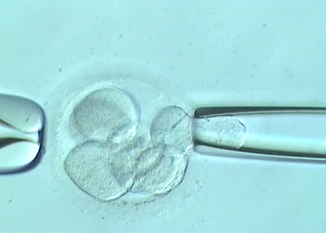
To explain the biopsy procedure we should keep in mind that our point of departure is EMBRYOS. Embryos are retrieved after performing an assisted reproduction cycle, preferably by Intracytoplasmic Sperm Injection (ICSI), and their development is assessed during the culture period until day 3 or day 5.
By day 3 biopsiable embryos should have a number of cells equal or higher than 6 so that their later development is not affected by the fact of having “captured” a cell to do the analysis. To perform the biopsy, a hole has to be drilled in the embryo’s zona pellucida (ZP), either by means of chemical agents (Tyrode’s acid) or laser pulses. A blastomere (an embryo cell) is then collected by aspiration and is subsequently analysed in our molecular and genetic biology laboratories.
Once the biopsy has been performed, embryos are kept under observation to assess their development until the time of transference.
By day 5 biopsiable embryos should have reached the blastocyst stage. If so, a minimum of 4-5 cells from the external cell layer that will form the placental structures (trophectoderm) are biopsied, keeping the inner cell mass that will form the embryo intact. Drilling of ZP is performed by laser, which is the most precise method available, as the thickness of the ZP is reduced by the associated processes of expansion, and the embryo’s development could be jeopardised by using other methods. The embryo will naturally extrude a piece of trophectoderm out of the zona pellucida. This is the piece that can be biopsied. The most common way to biopsy the trophectoderm is by aspiration, using laser pulses to trigger cell separation, although there are other methods.
Embryo biopsy is a delicate procedure that requires high precision and an experienced team of embryologists. Mistakes in performing the technique would jeopardise embryo development or the subsequent genetic diagnosis.
Dr. Dori Rodríguez, biologist at Instituto Bernabéu
To found out the next topics for our forum: follow us on facebook.
You can arrange an online consultation or book an appointment at Instituto Bernabeu.
More information on our website: www.institutobernabeu.com or www.ibbiotech.com