
What is the endometrium?
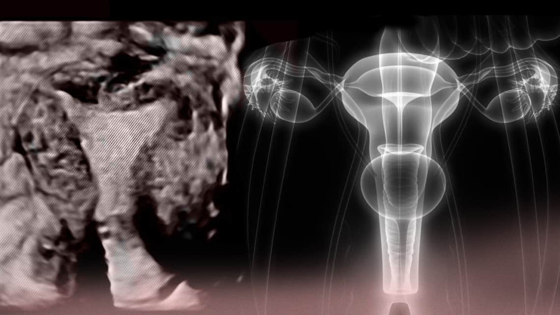
The uterus, the organ where pregnancy takes place, has a cavity inside where implantation occurs. This cavity is covered by a soft tissue called endometrium, the “nest” that the uterus prepares every month for a possible embryo.
Índice
Phases of devolopement of the endometrium
During the menstrual cycle the endometrium goes through 3 main phases:
- Menstrual phase, when it is shed in order to grow again.
- follicular phase or preovulatory, when it grows, and
- Lluteal phase or postovulatory when it reaches the proper state in which to produce implantation. These phases of the cycle are easily seen in an ultrasound.
1.- Menstrual phase
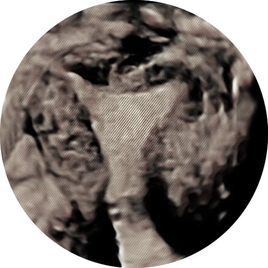
During the menstrual cycle we observe the endometrium as a thin white line (hyperechogenic) which can sometimes be seen mixed with menstrual blood (hypoechoic regions) along with uterine contractions that expel the menstruation. At this time the endometrium measures between 1-3 mm.
2.- Follicular phase
As we further progress in the menstrual cycle, we get to the follicular phase and in response to an ovarian stimulation a trilaminar pattern is seen. These are three parallel lines that grow 0.5mm per day, usually reaching a preovulatory size of between 9-14mm.
Three patterns are seen via ultrasound before ovulation:
- Type A. No triple line seen.
- Type B. A triple line is seen but without greater echogenicity in the internal line, so it is difficult to see.
- Type C. Triple line.
3.- Luteal phase
During the luteal phase, after ovulation, the progesterone produced makes the exterior lines thicker, so that they finally join with the interior line, making a single homogenous line at the start of the menstruation.
In Reproductive Medicine we pay a lot of attention to primarily the post ovulatory phase, since it can show reasons for implantation failure. It is important that the measurements and the visualization of the endometrial line should be done properly, and the sonographers experience as well as the machine used could play a role in incorrect diagnosis. Because of this the use of three dimensional ultrasound is quite promising. It allows us to not only evaluate the endometrial size on one plane, but the total endometrial volume and a study of the vascularization that feeds the endometrium, which is fundamental in uterine receptivity.
MORE RELATED INFORMATION:
- Recommendations following embryo transfer
- Implantation failure and repeated miscarriage. Treatment options
- Embryo transfer on day 3 or day 5. The pros and cons.
- The advantages and disadvantages of transferring embryos during a natural or artificial cycle
- Endometritis: What is it? How can it be detected? How can it be cured?
- Adenomyosis and recurrent implantation failure
Dra. Belén Moliner, gynecologist at Instituto Bernabeu.