
What can we see in ultrasound scans: follicles or oocytes?
Mistaking ovarian follicles for oocytes is one of the most common errors made by patients.
During ovarian stimulation, whether this is for regulating ovulation for insemination or for in vitro fertilisation, patients are administered different types of medicines depending on each case. However, there is one common aim: predicting and causing ovulation. This can be just once for patients attempting managed sexual intercourse and some types of insemination, or multiple for patients undergoing in vitro fertilisation treatment.
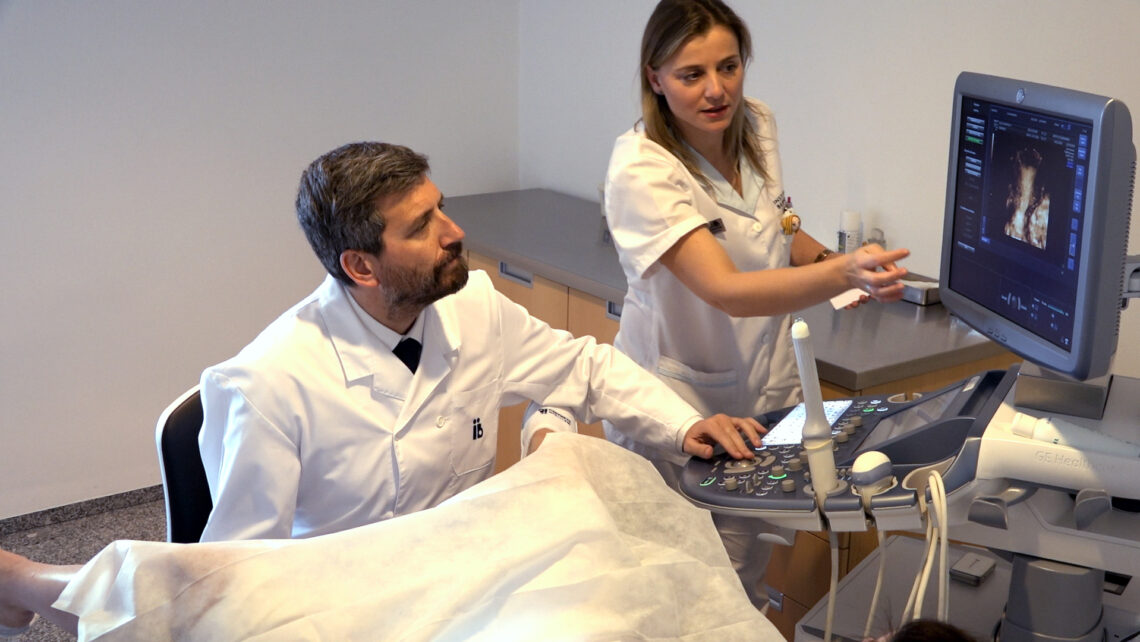
Ultrasound scans enable us to observe follicle development. The follicle is a structure that is made up of thousands of cells and it generally contains an oocyte.
The follicle may or may not contain an oocyte and the oocyte can be abnormal or normal, mature or immature. It is NOT UNTIL AFTER FOLLICULAR ASPIRATION THAT WE CAN ACTUALLY SEE THE OOCYTE ITSELF.
The oocyte is a cell and, therefore, it is microscopic. This is why it needs to be viewed in a laboratory.
Sometimes patients believe that the number of follicles is an indication of the number of oocytes. However, this is not always the case. Indeed, there are certain pathologies in the ovaries such as empty follicle syndrome (the name says it all) when follicles do not contain an oocyte.
Likewise, patients with poor ovarian response can have a greater number of follicles without oocytes or they can be abnormal oocytes, just like patients who suffer from implantation failure.
Last of all, it should be highlighted that the tests that we use in order to assess ovarian response – anti-Mullerian hormone levels and basal ultrasound scans – are indicators that do give us an idea but that do not give an exact prediction of the quantity and quality of the oocytes.
Dr Rafael Bernabeu, Medical Director at Instituto Bernabeu.