Røntgen i 3D-4D
A perfect picture of the foetus, almost like a photograph, it provides gynaecologists with reliable information for detecting a large number of pathologies.

What is three-dimensional ultrasound?
Three-dimensional ultrasound is the latest technology applied to ultrasound.Ultrasound allows us to navigate through the three spatial planes obtaining volumes of anatomical structures.
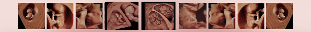
When applied to obstetrics, tridimension (3D) visualisation of the fetus allows a close approximation to what might correspond to a photograph showing with great clarity and realism the face and all body parts. When activated the cuatridimensión (4D), i.e., the time, there is foetal movement in real time and even all their facial expressions can be seen.
Advantages of 3D and 4D
This ultrasound examination system has many advantages over the conventional bidimensional scan:
- The images can be processed, stored and recovered in an instant.
- This therefore reduces scan times, since the image can be analysed by the physician once the patient has gone.
- The images can be saved and retrieved at the desired time. It can also be transferred to any computer and even sent within a network or via the Internet.
- This allows for the diagnosis to be compared and confirmed by colleagues who are present or connected to the network. A patient’s case can be studied in real time or on a deferred basis anywhere in the world by different experts.
- The technique also supports precise measurements of volumes of any part of the anatomy and the precise study of the vascularisation of all fetal organs.
Conditions for implementation
In order for 3D or 4D to be successful a number of requirements are essential:
- Right-foetal position. Logically, the foetal face is the most interesting structure searched in 3D: if the foetus has its back to the probe then it will be difficult to obtain the image. This Is easily solved using slight movements of the probe on the maternal abdomen.
- Foetal-Kinetics. The continuous movement of the foetus may be desirable in 4D, but in 3D it can make obtaining good images difficult. It is however a question of waiting a few moments and taking the best images when there is no foetal movement.
- At the time of exploration there should be abundant amniotic fluid surrounding the foetal surface you want to study. It may be necessary to mobilise the probe or even the mother in order to get the fluid to surround the fetal face.
- Proper calibrating of ultrasound equipment. It is indisputable that the equipment must be of the highest quality and the user must have had some training in its use. The device should be set according to the characteristics of the mother and the foetal position in order to obtain the best quality images at all times.
Benefits of the 4D Ultrasound
Three-dimensional ultrasound (3D) is a technique that goes beyond the diagnostic potential of ultrasound, especially in the field of Obstetrics. This situation has shown to be true in the following situations:
- Improving the diagnosis of many malformations, especially facial ones, size and foetal limbs. Greatly facilitates the parental understanding.
- There is an ability to observe, measure and evaluate the behavior of the foetus and its general movements in a more comprehensive comparison with conventional ultrasound. The four-dimensional ultrasound provides real-time foetal expressions (yawning, swallowing, sucking, grimacing, smiling, blinking) and various foetal movements (stretching, bending of head, isolated movements of arms and legs, etc.). It therefore allows us to study foetal neurology. It is known that foetal behavioral patterns directly correlate with the development of the central nervous system and the quality and intensity of foetal movements reveal its integrity. 4D ultrasound is therefore an indispensable tool for early detection of brain dysfunction. The ultimate goal is early detection of possible foetal distress or abnormal behavior before birth, not detected so far with prior examinations.
- There are contrasting advantages of a psychological nature. The mother can finally clearly see their child-to-be: there is always a great emotional impact that accompanies her until the end of pregnancy. It’s a new link that will connect the mother more to the child, to be able to live positively to the end of pregnancy with more security and confidence.