Endoscopic surgery
Endoscopy is defined as the direct observation of the inside of an organ or cavity by means of optical apparatice or endoscope (illumination tube connected to a video camera and television device), which is introduced through the natural orifices or through the abdominal wall. Thus we obtain information in a more reliable way than the type of information given by other external examinations, such as physical tests, ultrasound scans or radiology techniques.
By using endoscopy, we assure a clear interpretation of the images, minimizing the risk of complications. Endoscopic techniques allow the correct diagnosis and treatment of gynaecological pathologies in a less aggressive way, which is extremely important in the surgical treatment of reproductive problems.
The patient need only be at the clinic for a few hours, allowing them to go back home the same day of surgery. The post-operative feelings of discomfort are lower than with conventional surgery, which means that the patient does not have to modify substantially their daily activities.
Endoscopic surgery also provokes a lower risk of haemorrhaging and blood loss, and at the same time reduces adhesions. Besides, the aesthetic results with endoscopic techniques are much better that with conventional open surgery.
It is for these reasons that endoscopic surgery is recommended as being a reliable and safe technique.
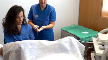
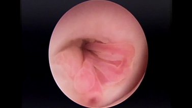
A diagnostic hysteroscopy consists of the introduction of a small-bore optic through the vagina and neck of the uterus. With the help of the infusion of physiological serum, the endometrial cavity is expanded, achieving good visualisation.
It also allows the taking of selective biopsy under direct visual control if it is considered appropriate. Patients do not need to be hospitalized and the intervention is carried out without anaesthesia or sedation being necessary. The procedure takes less than 5-10 minutes and once completed, patients can go back to their daily routine.
Since this procedure can cause feelings of discomfort, similar to menstrual pain, a previous preparation with muscle relaxant and analgesics is given.
Ambulatory diagnostic hysteroscopy can be used for the following:
- Suspicion of uterine adhesion.
- Suspicion of endometrial polyps.
- Suspicion of submocous myomas.
- Suspicion of uterine septums.
- Extraction of strange substances and IUD.
- Infertility research and implantation failures.
- Investigation of menstrual alterations.
- Study of endometrial cavity prior to assisted reproductive treatments.
This procedure requires general or local anaesthesia (sedation) as it is necessary to expand the neck of the uterus; that is why a pre-operative study and assessment is conducted by our anaesthesiologist.
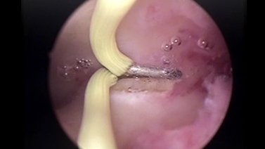
A surgical hysteroscopy solves most of the pathologies diagnosed by the ambulatory hysteroscopy and those found through other imaging techniques (ultrasound scan, hystero-salpingography, hystero-sonography, NMR, etc.).
Technically, it consists of introducing an optical cable and a surgical tool at the same time through the opening to the cervix, and this allows the use of small scalpels, coagulation systems (roller), etc.
Very few complications can occur with this procedure and, in general, are rarely serious. It is recognised as a very safe technique and a satisfactory way of dealing with intrauterine problems. It is usually possible for the patient to go back home the same day as surgery. Strenuous physical activity, swimming and sexual intercourse are not recommended for the following period of 5-7 days.
Surgical hysteroscopy is recommended for the following:
- Septoplasties.
- Myomectomies.
- Polypectomies.
- Ablation- endometrial reduction.
- Release of adhesions.
A laparoscopy is carried out under general anaesthesia, and therefore a preoperative study and assessment is required by our anaesthesiologist. The procedure consists of the introduction of CO² in the abdominal cavity by means of a needle inserted through the abdomen at navel level.
Once the distension of the abdominal cavity is achieved, the trocars or guides of 5 to 10mm are introduced, through which the surgical tools are located (optics, pressure forceps, scissors, coagulators, etc).
In a fertility study a uterine movilizator is placed in the inside of the uterus via the vagina. By means of the movilizator a methylene blue contrast is introduced in order to confirm the permeability of the Fallopian tubes.
The objective of the laparoscopy is twofold: on the one hand it is a tool for diagnosis and on the other; it can be used to solve the problems detected during surgery.
A laparoscopy can be used for:
- Fertility study.
- Diagnosis of uterine malformations.
- Unexplained pain or pelvic algia.
- Bilateral tubal sterilisation (tubal ligation).
- Endometriosis.
- Cysts and ovarian mass.
- Ectopic pregnancy.
- Ooforectomy (ovary extirpation).
- Myomectomy (myoma extirpation).
- Salpingectomy (extirpation of the pathological tubes).
- Ovarian drilling (polycystic ovary).
- Hysterectomy (uterus extirpation).
After 48-72 hours patients can go back to their daily routine.