What does an embryo biopsy entail?
Pre-implantation genetic diagnosis (PGD) is a technique that provides a ‘genetic understanding’ of the embryo before it is transferred to the uterus. Thanks to this technique, we are able to study the embryo’s chromosomal make-up and determine if it is a carrier of a hereditary condition of any kind. This information helps us to select the embryos that will lead to the birth of a healthy child. But how do we obtain this information?
Work is currently being carried out to discover non-invasive means of gathering genetic information from the embryo but, to date, the only means is an embryo biopsy. What is an embryo biopsy?
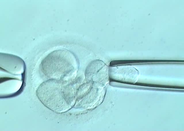
In order to explain the biopsy procedure, we need to take into account that THE EMBRYO is our starting point. The embryo is obtained following a cycle of assisted reproduction, preferably using intracytoplasmic sperm injection (ICSI). Development is evaluated during culture up until day 3 or 5. Embryo biopsies have traditionally been carried out on day 3. Nowadays, embryo biopsy on day 5 is the preferred procedure because it does not compromise embryo viability.
An embryo that can undergo a biopsy on day 3 needs to have 6 or more cells so that posterior development is not compromised as a result of having ‘captured’ a cell for analysis purposes. In order to carry out the biopsy, a hole is made in the embryo’s zona pellucida (ZP). Chemical agents (acid Tyrode’s solution) or laser pulses can be used in order to do so. Next, a blastomere (embryo cell) is obtained by means of aspiration and it is analysed in a molecular biology and genetics laboratory.
Once the biopsy has been carried out, the embryo is kept under observation and development is checked up until transfer.
An embryo that is to be biopsied on day 5 needs to have reached blastocyst stage. In these cases, a minimum of 4 to 5 cells from the external layer that forms the placenta structures (trophectoderm) are biopsied and the internal cell mass that leads to the embryo remains intact. The ZP is perforated using a more precise method involving laser because its thickness has decreased due to the expansion processes themselves. Other methods could damage the embryo and compromise development. The embryo naturally extrudes a fragment of the trophectoderm from the zona pellucida and this is the fragment that can be biopsied. The most common means of carrying out a biopsy on the trophectoderm is aspiration aided with laser pulses in order to separate the cells. Other methods, however, are available.
Embryo biopsy is a delicate procedure. It requires a lot of precision and an experienced team of embryologists because mistakes in the technique can compromise embryo development or posterior genetic diagnosis.
Dr Dori Rodríguez, biologist at Instituto Bernabeu.