
Pre-implantation genetic diagnosis (PGD): comprehensive chromosome screening
Pre-implantacional genetic diagnosis (PGD) is a technique used to detect genetic or chromosomal abnormalities and it is performed on the embryo prior to transfer to the mother’s uterus.
Its use is recommended in couples where one of the partners is a carrier of a genetic or chromosomal abnormality that either means they are unable to get pregnant or increases the risk of pregnancy loss or abnormalities in their children. In some cases, it is the only means of having biologically healthy children and, in others, it is an alternative to prenatal diagnosis.
Comprehensive chromosome screening (PGS/PGT-A/CCS) is a version of PGD . This technique is mainly recommended for two particular cases. In the first instance, couples who, despite not having a genetic or chromosomal abnormality, experience recurrent pregnancy losses during naturally-conceived pregnancies or following in vitro fertilisation. Secondly, couples who experience implantation failure. That is, couples who have not managed to get pregnant following several courses of IVF with good quality embryos and with no apparent cause. Both situations can occur because the embryos have chromosomal abnormalities and, as a result, do not implant or, if they do implant, lead to spontaneous pregancy loss shortly after implantation.
Patients who have experienced an aneuploidy in a previous pregnancy, those who are at a greater risk due to age (women over 35 years of age) and couples where the male partner has a high aneuploidy rate in spermatozoa (abnormal FISH in spermatozoa) can also benefit from this technique.
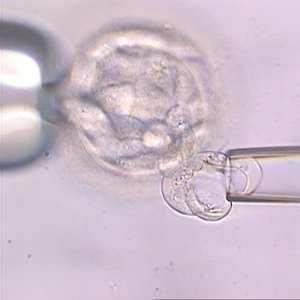
Couples have to go through assisted reproduction treatment (IVF/ICSI) in order for the technique to be carried out. Once the oocyte has been fertilised with the sperm in the laboratory, the embryos are left to cultivate until they reach blastocyst stage (days 5, 6 or 7 of embryo development) and between five and ten cells are removed from the outer cell layer of the embryo (trophectoderm). This procedure is called embryo biopsy. Once embryo biopsy has been completed, genetic analysis of the cells is carried out and the healthy embryos that can be transferred to the uterus are diagnosed.
Chromosome analysis (PGS/PGT-A/CCS) consists of analysing the 23 pairs of chromosomes in the embryo. Embryos with the normal number of chromosomes (euploid) have a greater chance of implanting and leading to both a pregnancy and the birth of a healthy child compared with embryos that have chromosomal abnormalities. Therefore, by selecting and transferring only chromosomally-normal embryos, this technique increases the chances of having a healthy child.
There is often a fear that removing cells from the embryo will have a negative impact on the future baby but it is important to point out that removing some cells from the outer cell layer of the embryo (the future placenta) when it has thousands of cells affects neither implantation nor normal division and, therefore, poses no risk to the development of the future foetus.
Dr Ruth Morales, molecular biologist at Instituto Bernabeu.