
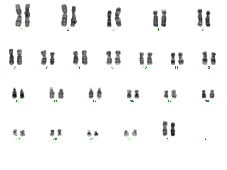
Genetic karyotype
All our genetic information is encoded in DNA. DNA is found in the nucleus of every cell of our body as part of the structures called chromosomes. The analysis of these chromosomes is called a karyotype.
A karyotype is performed on cells called lymphocytes obtained from a simple blood sample, but it can also be performed on amniotic fluid cells from amniocentesis or chorionic villi.
Each chromosome can be distinguished from the rest by its shape, size and banding pattern which can be observed along the entire chromosome after specific staining completed in the laboratory.
The alterations detected in a karyotype can be classified into two categories: Numerical alterations and structural changes.
1. Numerical alterations:
The number of chromosomes is characteristic of each species. Human beings have 46 chromosomes (23 pairs) in the nucleus of every cell in our bodies. These 23 pairs are arranged in 22 pairs, called autosomes, and a pair of sex chromosomes (X and Y) which differentiates the sexes. Women have two X chromosomes, whereas men have one X and one Y chromosome.
The most widely-known numerical alteration is Down’s syndrome. In the karyotypes of these patients there are three chromosome 21 instead of the usual two, causing varying degrees of learning disabilities.
There are other numerical alterations that generate reproductive problems. For example, men with Klinefelter syndrome have two X chromosomes and one Y. These patients suffer testicular failure resulting in fertility problems.
On the other hand, women who suffer from Turner syndrome have only one X chromosome karyotype. The absence of one X chromosome causes underdeveloped sexual characteristics and consequently leads to infertility.
2. Structural alterations:
Certain chromosomal regions are found to be duplicated or even absent. This gain or loss of genetic material has extremely varied consequences depending on the genes involved.
Other structural abnormalities are translocations and inversions. In the first case there is an exchange of material between two chromosomes and in the second, a chromosomal region is inverted with respect to its normal position.

Structural alterations may have different clinical consequences such as low semen quality in men, low ovarian reserve and repeat miscarriage in women.
If a chromosomal abnormality is detected in one member of the couple, the solution to ensure healthy biological descent is Preimplantation Genetic Diagnosis (PGD).
PGT-SR is a PGD (Preimplantation Genetic Diagnosis) variation technique for detecting chromosomal alterations in an embryo prior its transfer to the mother’s uterus.
In addition to the alterations already mentioned, there are chromosomal variants called polymorphisms. These polymorphisms are relatively common in the general population and show no apparent phenotypic consequences, but recent scientific studies have linked them to infertility.
A karyotype is an important diagnostic test in patients who require assisted reproduction techniques because there is a higher incidence of abnormal karyotype in couples with fertility problems. This technique is indicated mainly in cases of seminal alteration, ovarian failure and in couples with repeat miscarriage or implantation failures.
There’s another technique called array CHG (aCGH) which detects chromosomic alterations (small chromosomal regions duplicities or absences) with a sensibility level much higher than conventional Karyotype. Approximately, the aCGH reaches diagnosis levels 10 times higher than karyotype. This technique is a big step forward in cases with unknown infertility and recurrent implantation failure (know more about Array-CGH).
Dr José Antonio Ortiz, Molecular Biologist at Instituto Bernabeu.